When the mucous membrane is removed the surface of the cartilage is seen to be indented by a number of small pits in which mucous glands are lodged. The posterior commissure of the larynx is a name often given to the posterior portion of the glottis.

Posterior View Of Larynx Human Body Vocabulary Medical Knowledge Medical Anatomy
F upper ring of the windpipe.

. Front view A epiglottis. Get premium high resolution news photos at Getty Images. There is a printable worksheet available for download here so.
The inferior aspect of the. Maybe you have knowledge that people have search numerous times. The larynx houses the vocal cords and manipulates pitch and volume which is essential for.
Anatomy Of The Larynx Posterior View. PHARYNX AND LARYNX First click on Systems then under Respiratory System Views click on 4. The longer superior horn along with the entire superior border of the thyroid cartilage attaches to the hyoid bone by the thyrohyoid membrane.
There is a printable worksheet. It is narrower towards the front and wider in the back with a midline ridge that serves as a point of attachment for the esophagus. Gemmellposts 1 Comment Although the person is speaking in German in the following video it is fun to see how of parts of your body work together to create speech and in a more sustained way singing.
Cartilages and Ligaments of the Larynx. This is an online quiz called Anterior and Posterior view of Larynx. Pass from muscular process of one to the apex of the opposite arytenoid.
The true folds meet anteriorly at the anterior commissure small arrow. Its lower part projects backward as an elevation the tubercle or cushion. It draws arythenoid nearer to each other and adduct the vocal folds.
The Anatomy of the Larynx. No food or drink shall pass into his presence without dire consequences Buy this organ and its activity pages by following the links below. Hyoid bone Epiglottic cartilage Arytenoid Cricoid cartilage Thyrohyoid ligament cartilage Thyroid cartilage Cricotracheal ligament Trachea Cricothyroid ligament Corniculate.
Attach opposite posterior surface of the arytenoid cartilage. The true vocal fold 1 extends from anterior to posterior and is separated from the false vocal fold 2 by the ventricle arrowheads. Crosses each other on the posterior surface of the transverse arythenoid.
CT can define the anatomy of the postoperative larynx and can assist laryngoscopy in the detection of recurrent neoplasm Fig. Posterior view of the larynx. This game is part of a tournament.
About this Quiz. The superior aspect of the cavity laryngeal inlet opens into the pharynx inferior and posterior to the tongue. There are many muscles that either make up a certain part of the laryngeal structure inside the neck or that sit adjacent to it and aid in its functionThese muscles produce the movements of the larynx and its cartilages thus enabling the proper air conduction speech movements of the epiglottis and airways protection.
The interarytenoid muscles are part of this anatomical landmark. It consists of elastic cartilage and gives flexibility to the epiglottis which is a heart-shaped cartilage covered with mucous membrane. Colouring Activity Pages A5 JPEG.
The posterior margin of each lamina extends upward into a superior horn and downward into an inferior horn. Label the posterior view of the larynx based on the hints if provided. 4 rows This is an online quiz called Posterior View of the Larynx.
Label the posterior view of the larynx based on the hints if provided. It represents the lower portion of the larynx. Posterior View of Lord Larynx ImagePosted on September 29 2017September 4 2018by thecomicalanatomist Meet the backside of Lord Larynx the producer of sound.
Explore the structures of the pharynx and larynx here specifically you can see. March 3 2017 Author. Label the posterior view of the larynx based on the hints if provided.
Physical examination of patients who have undergone prior irradiation or surgery may be difficult because of altered normal laryngeal anatomy and swelling of the adjacent soft tissues. You need to be a group member to play the tournament. The opening of larynx into pharynx known as the laryngeal inlet is about 45 centimeters in diameter.
The muscles of the. The posterior or laryngeal surface is smooth concave from side to side concavo-convex from above downward. Label the blood vessels of the female pelvis using the hints provided.
Sitting just below the thyroid cartilage the cricoid cartilage is ring-shaped and encircles the airway. Terminology The term commissure is a misnomer as the true vocal cords do not join together posteriorly to form a commissure 34. The image is rotated 180 degrees from the usual perspective of the endoscopist.
Click on Menu to return to the systems view now click on 6. This online quiz is called Posterior View of Larynx. The larynx ˈ l æ r ɪ ŋ k s commonly called the voice box is an organ in the top of the neck involved in breathing producing sound and protecting the trachea against food aspiration.
The form of the lateral aspects is determined by the larynx cartilages and consist of three parts a superior one that matches the thyroid cartilage an inferior one that matches the cricoid cartilage and a middle. Start studying Larynx Posterior View. Endoscopic view of the larynx using an office endoscope.
View the full answer. 3D anatomy tutorial on the cartilages of the larynx from AnatomyZone For more videos 3D models and notes visit. Learn vocabulary terms and more with flashcards games and other study tools.
Label the structures of the larynx anterior and posterior views by clicking and dragging the labels to the correct location. Muscles of the larynx. The epiglottic cartilage lies posterior to the root of the tongue and hyoid bone and anterior to the laryngeal inlet and it forms the superior part of the anterior wall and the superior margin of the inlet.
The posterior part of the internal space of the larynx is part of the anterior wall of the pharynx and has two vertical recesses referred to as the piriform sinus. The pharyngeal constrictors superior middle inferior.

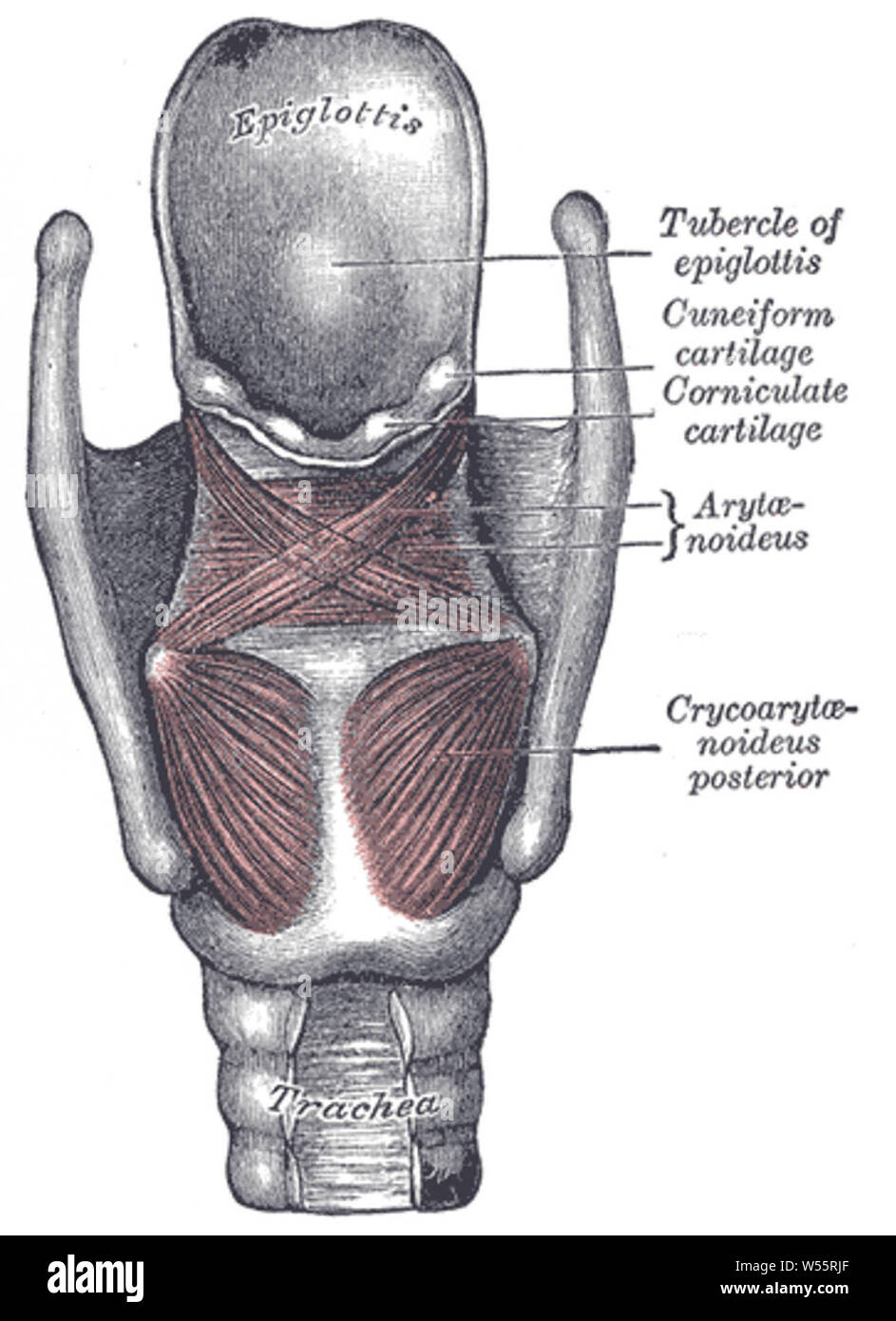
Posterior View Of The Larynx And Vocal Cords Bones Muscles Cartilages Stock Photo Alamy

Cartilages Of The Larynx Posterior View Diagram Quizlet

1 5 Posterior View Of Larynx Showing Aryepiglottic And Oblique Download Scientific Diagram
Posterior Larynx Anatomy With Annotations Wall Art Canvas Prints Framed Prints Wall Peels Great Big Canvas

Schematic Of The Human Larynx Framework Based On Gray 6 A Download Scientific Diagram
Muscles Of Larynx Posterior View Stock Photo Alamy

Larynx Anatomy With Labeled Structure Scheme And Educational Medical Views Anterior Posterior And Cross Section Examination With Trachea Parts Vector Illustration Vocal Cords Housing Description Royalty Free Cliparts Vectors And Stock Illustration

Larynx Anatomia Laringe Humor
0 komentar
Posting Komentar